AFib vs atrial flutter: how to tell them apart on the ECG
Both start in the atria, both can drive the heart fast, and they are easy to mix up on a monitor. The difference comes down to two things: whether the rhythm is regular, and what the baseline between the beats looks like.
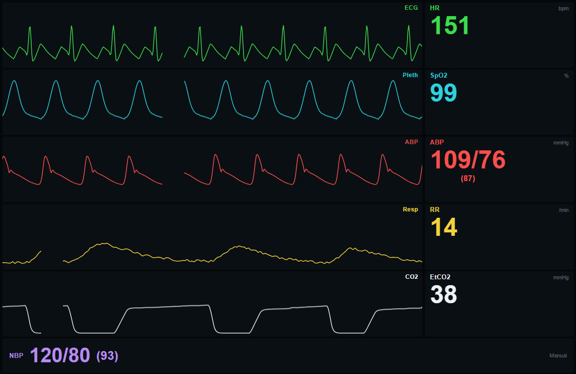
Watch it on a monitor
Both rhythms live on the monitor, ECG and pulse curves side by side.
Plays from YouTube when you click. Nothing loads from YouTube before that.
Atrial fibrillation: chaos in the atria
In atrial fibrillation the atria have no organised electrical activity at all. Hundreds of small, chaotic impulses a minute reach the AV node, which lets some of them through at random. On the ECG that gives:
- No real P waves, only an uneven, wavy baseline (fibrillatory waves) that can be coarse or so fine it looks almost flat.
- Irregularly irregular R-R intervals: no two gaps between beats are quite the same, and there is no pattern.
- Narrow QRS complexes, unless there is also a bundle branch block.

Atrial fibrillation, 6 seconds at 25 mm/s. Count the small squares between the R waves: they are never the same twice.
Atrial flutter: one circuit going round and round
Atrial flutter is organised. A single electrical circuit, usually in the right atrium, loops at roughly 300 times a minute. Each loop makes a flutter wave, and together they form the classic sawtooth, clearest in leads II, III and aVF. The AV node cannot pass on 300 impulses a minute, so it typically lets every second one through. That is 2:1 conduction, and it gives a ventricular rate of about 150.
- Sawtooth flutter waves instead of P waves, at about 300 a minute.
- Usually regular R-R intervals, because the block tends to be fixed, most often 2:1.
- A heart rate that is often very close to 150.

Atrial flutter with 2:1 conduction, 6 seconds. Every other flutter wave sits in or right next to the QRS and T wave, which is why 2:1 flutter is so easy to miss.
Four questions that separate them
- Is the rhythm regular? Irregularly irregular points to fibrillation. Regular points to flutter, or to something else entirely.
- What does the baseline look like? A chaotic, uneven wobble suggests fibrillation. A repeating sawtooth suggests flutter.
- Is the rate around 150? A regular narrow-complex tachycardia at almost exactly 150 should always make you think of 2:1 flutter.
- Is the block changing? Flutter with variable block, 2:1 mixed with 3:1 or 4:1, turns irregular and can pass for fibrillation. The R-R intervals then jump between a few fixed lengths rather than varying at random.
Why 2:1 flutter is so often missed
At 150 a minute there is not much room between the beats. One flutter wave lands just before the QRS and looks like a P wave, the other hides in the T wave. The strip then passes for sinus tachycardia or SVT. Looking at several leads helps, and so does anything that briefly slows conduction through the AV node, which clinicians sometimes do on purpose to uncover the flutter waves.
What the pulse curves show
The monitor gives another clue besides the ECG. In atrial fibrillation the beats come at random intervals, so the heart fills unevenly: a beat after a long pause pushes out more blood than one that comes early. The pleth and arterial curves therefore change height from beat to beat, and some short beats barely show (there is a picture of this in how to read a patient monitor). In 2:1 flutter the pulse waves are even, because the beats are.
Practising the difference
Rhythm recognition sticks through repetition. The simulator has both rhythms: atrial fibrillation with irregular R-R intervals and pulse waves that vary beat by beat, and atrial flutter with 2:1 conduction at 150. An instructor can switch between them, or from sinus rhythm into either, and ask the group what just happened. Every rhythm is described on the ECG simulator page.
Frequently asked questions
What is the main difference between AFib and atrial flutter on the ECG?
Atrial fibrillation is irregularly irregular, with no P waves and an uneven baseline. Atrial flutter shows regular sawtooth flutter waves at about 300 a minute and is often regular itself, typically at 150 with 2:1 conduction.
Why is the heart rate often 150 in atrial flutter?
The flutter circuit runs at around 300 a minute and the AV node usually passes every second impulse. Half of 300 is about 150.
Can atrial flutter be irregular?
Yes, if the block varies, for example between 2:1 and 4:1. The R-R intervals then switch between a few fixed lengths, unlike the completely random intervals of atrial fibrillation.
Is this medical advice?
No. This is a teaching article for practising ECG recognition. Diagnosis and treatment belong to qualified clinicians with the full clinical picture and a 12-lead ECG.
Last updated