EtCO2: what it means and what the normal values are
EtCO2, end-tidal carbon dioxide, is the amount of CO2 in the breath at the very end of exhalation. In a healthy adult it sits around 35 to 45 mmHg, or 4.7 to 6.0 kPa. What makes it useful is how quickly it moves when breathing or circulation changes.
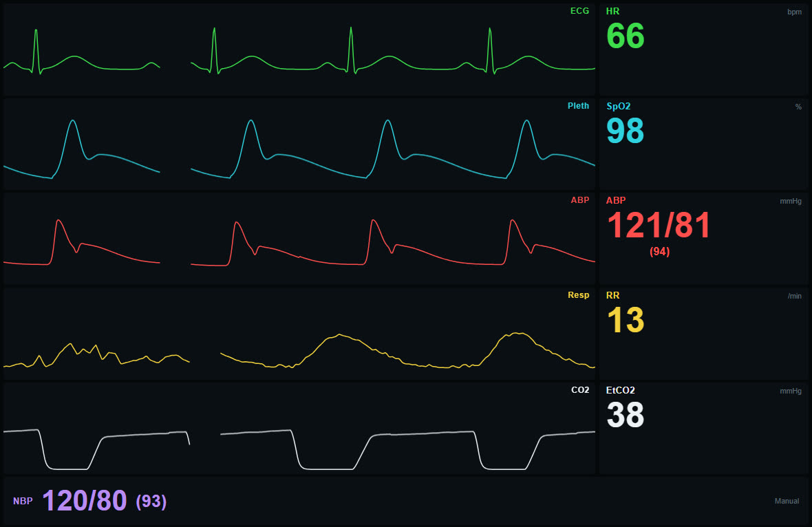
What EtCO2 measures
A sensor in the airway, or a thin sampling line in a nasal cannula, measures CO2 in the breath continuously. The curve it draws is the capnogram. The number is the highest value at the end of each breath, which is as close as you get to the gas that sat deep in the lungs.
Three things decide that number: how much CO2 the body produces, how much blood carries it to the lungs, and how well the lungs breathe it out. A change in any one of them shows up in EtCO2, which is why it is read together with the patient and the rest of the monitor rather than on its own.
Normal values
- Adults at rest: about 35 to 45 mmHg, which is 4.7 to 6.0 kPa.
- EtCO2 is normally a few mmHg (about 0.3 to 0.7 kPa) lower than the CO2 in an arterial blood gas.
- That gap widens when parts of the lung are ventilated but get little blood flow, for example with a pulmonary embolism, low cardiac output or severe lung disease. EtCO2 then underestimates the real CO2 level.
What a low EtCO2 means
- Breathing too much: anxiety, pain, or being bagged too fast.
- Poor circulation: shock, heart failure, pulmonary embolism, cardiac arrest. Less blood reaches the lungs, so less CO2 is breathed out even if ventilation is normal.
- A sudden drop to zero: think of the equipment and the airway first. A disconnected circuit, a tube that has slipped out or into the oesophagus, a blocked airway or apnoea.
What a high EtCO2 means
- Breathing too little: opioids, sedation, exhaustion, or a ventilator set too low.
- CO2 retention: some patients with severe COPD live with a high level.
- More CO2 produced: fever, shivering, sepsis, and rarely malignant hyperthermia.
- Rebreathing: if the baseline no longer returns to zero between breaths, the patient is breathing in CO2. The shape of the curve shows this better than the number, see reading a capnogram.
EtCO2 during CPR
During cardiac arrest the lungs only see the blood flow that chest compressions create, so EtCO2 becomes a rough measure of how good the compressions are. Very low values suggest ineffective compressions or a tired compressor. Targets commonly cited are above roughly 10 to 20 mmHg (1.3 to 2.7 kPa), and local guidelines differ. A sudden, sustained jump, often towards normal values, can be the first sign of return of spontaneous circulation, sometimes before anyone feels a pulse.
kPa or mmHg?
It is the same measurement in two units. The Nordic countries mostly use kPa, while many other countries use mmHg. One kPa is about 7.5 mmHg, so 5.0 kPa is roughly 38 mmHg. This simulator shows EtCO2 in mmHg on the English, Spanish, French and German versions and in kPa on the Swedish, Danish, Finnish and Norwegian ones.
Practising with the simulator
The instructor sets EtCO2 with its own slider, and the capnogram follows with realistic angles and a plateau that rises slightly through each breath. In apnoea and cardiac arrest rhythms the curve goes flat at zero and the number shows no reading, because there is no breath to measure. EtCO2 is deliberately not tied to blood pressure, so the instructor can show poor compressions, a sudden rise at ROSC, or a patient slowly retaining CO2. More on the EtCO2 simulator page.
Frequently asked questions
What is a normal EtCO2?
About 35 to 45 mmHg in adults, which is 4.7 to 6.0 kPa.
What does a low EtCO2 mean?
Either the patient is breathing too much, or too little blood is reaching the lungs, as in shock, pulmonary embolism or cardiac arrest. A sudden drop to zero usually means a problem with the airway or the equipment.
Is EtCO2 the same as PaCO2?
No. EtCO2 is measured in the breath and is normally a few mmHg lower than PaCO2 in an arterial blood gas. The gap grows with pulmonary embolism, low cardiac output and lung disease.
Last updated