Why PEA confuses new nursing students, and how to teach it properly
A monitor showing a normal-looking heart rate on a patient with no pulse is the single hardest thing to teach from a screen, because the mistake it corrects for is invisible until someone checks a pulse.
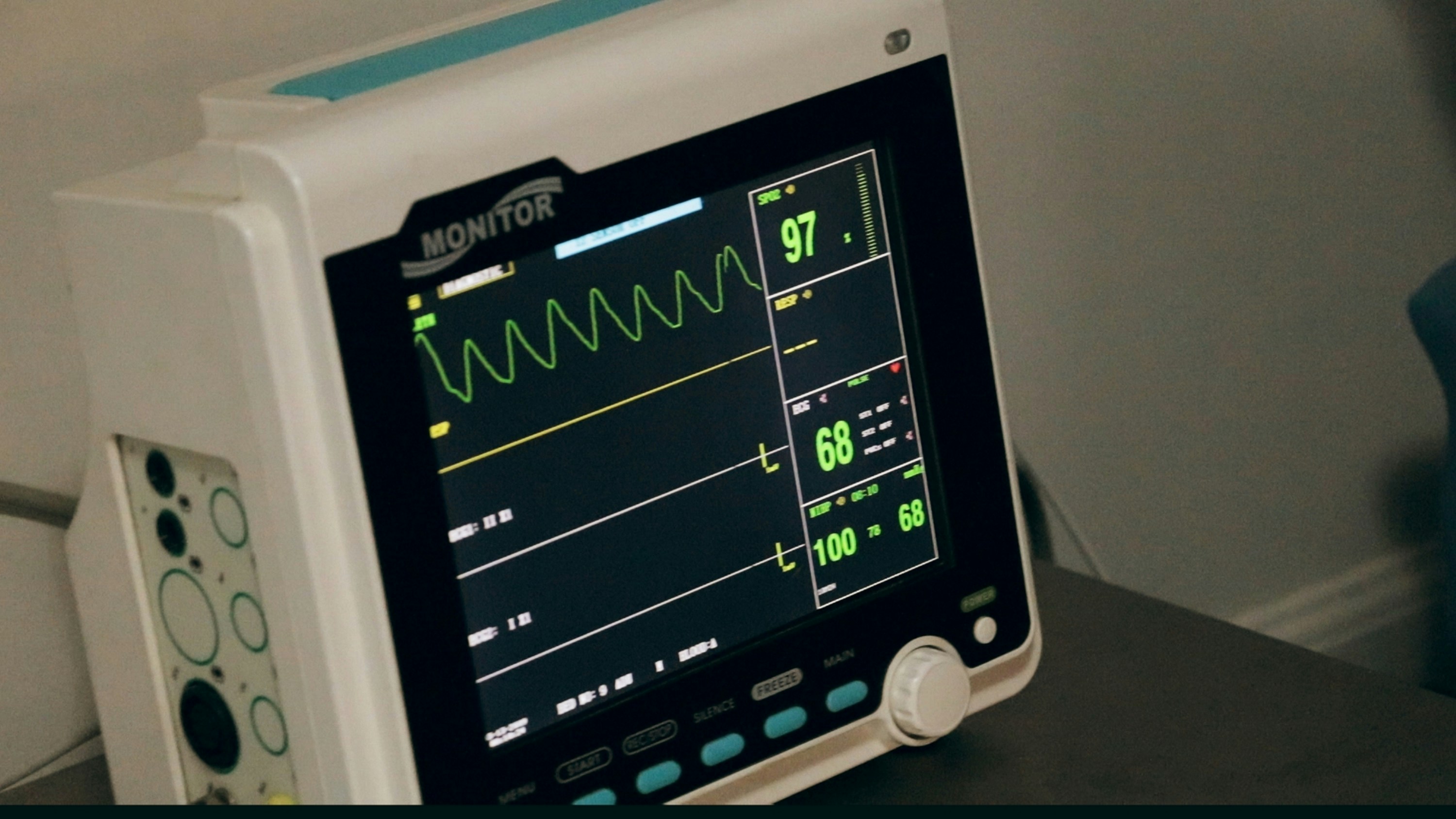
PEA is a diagnosis you can't make from the screen
Pulseless electrical activity is exactly what the name says: organized electrical activity on the ECG, at a rate that can look entirely normal, in a patient who has no pulse. That's the part new students find hard to accept. A strip with a clean QRS complex marching along at 80 beats a minute looks like a stable patient, right up until someone puts two fingers on a carotid artery and finds nothing there. The ECG can't tell you PEA is happening. Only a pulse check can.
Why students reach for asystole instead
Ask a nursing student what a cardiac arrest rhythm looks like and most of them will draw a flat line. That's asystole, and it's the rhythm that matches the word "arrest" in their heads: no electrical activity, no pulse, unmistakable on a strip. PEA breaks that mental model completely, because the strip looks fine. Students who have only ever practiced recognizing rhythms by eye, not by combining a rhythm with a clinical check, will read a normal-looking QRS complex as a normal-looking patient. The gap between what the ECG shows and what the patient's circulation is actually doing is the entire teaching point, and it's also exactly the gap a slide deck can't close.
The setup mistake that gives the diagnosis away
The easiest way to ruin a PEA scenario is to make the patient look unwell in every other respect at the same moment the rhythm would otherwise look normal. A patient who is unresponsive, gray, and not breathing, with a monitor reading a heart rate of 78, doesn't test whether a student can catch PEA. It tests whether they can catch "this scenario is obviously staged." A believable PEA scenario needs to hold the rest of the picture close to normal for long enough that the pulse check is the thing that actually reveals the problem, not a formality after every other sign has already spelled it out.
Building a scenario that actually tests for it
Start the patient stable, with a rhythm and a heart rate that would pass for a normal round. Let the trainee begin their assessment on that baseline. Partway through, without changing the monitor's rate or rhythm at all, script the moment where the patient becomes unresponsive, or where a colleague reports no pulse felt on a routine check. The rhythm on the display doesn't need to change for the scenario to work, and in a real PEA arrest it often doesn't. What has to change is what's expected of the trainee: they either check a pulse on their own initiative, at the right moment, or they don't, and reading the same organized rhythm as "the patient is fine" is the exact failure the exercise is designed to catch.
On patientmonitorsimulator, that means picking one of the organized rhythms, sinus, SVT, even a rhythm with a wide QRS, and leaving the heart-rate slider wherever it started. The screen never labels what's happening, so nothing on it contradicts the story you're telling as the instructor. The teaching moment lives entirely in the gap between the monitor and the patient, exactly where it lives in a real arrest.
Debrief questions that test the gap, not the rhythm strip
A debrief that only asks "what rhythm was that?" rewards students for reading a strip, which was never the hard part of a PEA scenario. Better questions target the moment the student should have gone looking for information the monitor can't provide: When did you last confirm a pulse? What would have told you the heart rate on the screen wasn't the whole story? What's different about how you'd manage this compared to a stable patient with the same rhythm? Those questions push the discussion toward the clinical habit the scenario is meant to build, checking a pulse rather than trusting a number, instead of toward rhythm identification the strip already gave away for free.
Where PEA and asystole sit next to the other arrest rhythms
Ventricular fibrillation and pulseless VT are the two rhythms where the strip itself is the emergency, chaotic or fast enough that no clinician needs a pulse check to know something is badly wrong. Asystole sits at the other extreme: a flat line that announces itself. PEA is the awkward middle case, a rhythm that offers no visual clue at all, which is exactly why it's worth a training scenario of its own rather than folding it into a general "recognize the arrest rhythms" drill. Students who can already name all four arrest rhythms on a static strip still get caught out by PEA the first time it's embedded in a scenario instead of printed on a quiz.
Frequently asked questions
What's the difference between PEA and asystole?
Asystole is no electrical activity at all, a flat line. PEA is organized electrical activity, sometimes at a completely normal-looking rate, in a patient who has no pulse. The ECG alone can't distinguish PEA from a stable rhythm; only a pulse check can.
Why is PEA hard to simulate on a monitor?
Because the monitor is supposed to look normal. A good PEA scenario keeps the rhythm and heart rate unchanged and moves the teaching moment into the pulse check itself, rather than into anything visible on the screen.
Can patientmonitorsimulator run a PEA scenario?
Yes. Pick any organized rhythm and hold the heart-rate slider steady, then script the patient becoming unresponsive or a pulse check coming back negative without changing anything on the display.
Last updated